

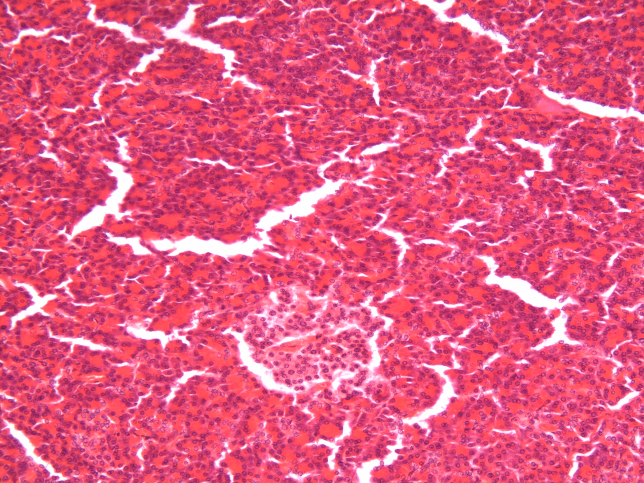
The pancreatic islets (indicated by the black circles) appear as "islands" of lighter stained cells in a "sea" of more darkly stained acini ("wine") cells. The tissue cracked during slide prep. Not all slides have the cracking seen in these photos.
Photo taken by Dr. W. Coons (100x)
The pancreatic islets (indicated by the black circles) appear as "islands"
of lighter stained cells in a "sea" of more darkly stained acini ("wine")
cells. The tissue cracked during slide prep. Not all slides
have the cracking seen in these photos.
Photo taken by Dr. W. Coons (200x)

The pancreatic islets (indicated by the black circles) appear as "islands"
of lighter stained cells in a "sea" of more darkly stained acini ("wine")
cells. The tissue cracked during slide prep. Not all slides
have the cracking seen in these photos.
Photo taken by Dr. W. Coons (400x)


The pancreatic islets (indicated by the black circles) appear as "islands"
of lighter stained cells in a "sea" of more darkly stained acini ("wine")
cells. The tissue cracked during slide prep. Not all slides
have the cracking seen in these photos.
THYROID GLAND
PARATHYROID
GLAND
PANCREAS
ADRENAL GLAND
(SUPRARENAL)
TESTIS
OVARY
PITUITARY GLAND
(HYPOPHYSIS)
Back to Index Page Back to Course Supplements Back to VC Homepage